
1888–1920: from Nebraska to a workshop in San Diego
Rife was born in 1888 in Elkhorn, Nebraska. In 1913, at the age of twenty-five, he arrived in San Diego after driving across the country from New York. Lynes introduces him as a happily married man drawn to machines, optics, electronics, and bacteriology. His friends called him Roy and emphasised his gentleness, modesty, and exceptional skill.
His interests ranged from ballistics and building racing cars to photography and microscopy. In its account of these early years, the book also mentions service in the Navy during the First World War and visits to European laboratories on behalf of the American government. Rife was acquiring the knowledge that allowed him to turn a research idea into a precisely engineered instrument himself.
He began building his first microscope in 1917 and completed it in 1920. His first radio-frequency instrument also dates from that time. While observing organisms, he became interested in whether their different chemical and electrical properties also meant that they responded differently to electrical frequencies. His experiments required microscopes, tubes, electronic components, photographic equipment, laboratory cultures, and animals. The industrialists Timken and Bridges funded the laboratory and his early research.
A colleague remembers Rife at work
Lynes brings the nature of his work to life through a colleague’s recollection from 1958. It describes a man who made his own microscopes, a micromanipulator (a device for moving tiny objects very precisely under a microscope), and tiny surgical instruments, operated carefully on experimental animals, and spent long hours sitting motionless over specimens. The colleague also remembered the orderly laboratory and Rife’s patience. This portrait helps explain why his associates saw him as much more than the inventor of a single device. [1]
The twenties: the desire to see life
The first area the book describes in greater detail is tuberculosis. Rife tried to identify the electrical response of its causative organism and observe the effect under the microscope. According to Lynes, he succeeded in destroying the bacterial forms he could see, yet some of the experimental animals still died. Rife concluded that a smaller form, invisible to the eye, remained. To investigate it, he needed a different way of observing.
Between 1920 and 1925, approximately twenty thousand sectioned and stained tissue samples were prepared. Even so, he did not find what he was seeking. He believed that chemical dyes and specimen preparation altered or destroyed tiny structures before the researcher could examine them. Out of this dissatisfaction grew the idea that Lynes calls “staining with light”: a microorganism would become visible under suitable illumination, without conventional chemical staining.
He completed his second microscope in 1929. A front-page article in the San Diego Union on 3 November described his instrument and his search for a faster way of preparing living specimens. Rife explained to the journalist the advantage of a researcher who was also a mechanic: he understood the question and could build the tool needed to answer it. A later newspaper account compared his approach with astronomy, in which an observer identifies celestial bodies by their light.
In the chapter on the twenties, Lynes dates the start of Rife’s cancer research to 1922. For Rife, the twenties were a period of sustained work that he himself called pure science. With the news coverage in 1929, his solitude ended: visitors, letters, requests for demonstrations, and offers of collaboration began to arrive. At the same time, the Great Depression began. Lynes sees this as a transition from relatively independent research to a decade in which Rife would have to deal not only with technical questions but also with money, reputation, and influence. [1]
1931: meeting Kendall and Johnson
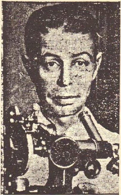
Two crucial supporters entered Rife’s life in 1931. Arthur I. Kendall headed medical research at Northwestern University in Chicago. Milbank Johnson was a physician, a member of a Pasadena hospital’s governing body, and an influential figure connecting medical circles in southern California. Kendall had heard about the unusual microscope and asked Johnson to investigate the reports.
Johnson visited Rife with the pathologist Alvin G. Foord and two other physicians. He reported what he had seen to Kendall, who arrived in California in mid-November. Kendall brought his culture medium, K Medium. He used it to study forms of bacteria small enough to pass through filters that retained ordinary bacteria. Now he wanted to see what he had been detecting through cultures and filtration.
The book describes an experiment with the organism that causes typhoid fever. After culturing and repeated filtration, the two researchers observed tiny, moving, turquoise-blue bodies under Rife’s microscope. Lynes reports eighteen repetitions of the test. In the optical explanation presented, controlled polarised illumination (illumination using light whose oscillations have been oriented in a particular way) was essential: adjusting the prisms was said to produce a strong colour contrast between the observed bodies and the colourless background of the medium.
The dinner, newspaper reports and publications of 1931
On 20 November, Johnson held a dinner in honour of Kendall and Rife. More than thirty physicians, pathologists, and bacteriologists attended. The Los Angeles Times reported on the event two days later and subsequently published a photograph of the two researchers. The newspaper account summarised by Lynes attributed a working magnification of 5,000 to 17,000 times to the microscope and emphasised its new arrangement of lenses, double quartz prisms, and powerful illumination.
In December, their paper Observations on Bacillus Typhosus in its Filtrable State appeared in California and Western Medicine. On 11 December, the news supplement of Science published an item about filterable bodies visible with Rife’s microscope. At the end of the month, a newspaper also reported a presentation to two hundred and fifty scientists. Lynes portrays 1931 as the moment when the new optics and Kendall’s cultures seemed capable of opening a broader direction for research. [2]
1932: a dispute over bacteria, viruses, and observation
To explain the significance of Kendall’s experiments, Lynes takes the story back to the dispute between Béchamp and Pasteur and to pleomorphism, the changing forms of microorganisms. In his interpretation, medicine adopted too rigid a distinction between bacterium and virus and overlooked researchers who linked different forms within a single life cycle. He places Rife’s work within this longer research tradition, which he believes was neglected.
Béchamp’s view of changes in microorganisms
Lynes describes Béchamp’s explanation in more detail: the change supposedly concerned the size of bacteria as well as their shape. Béchamp called the tiny particles microzymas. He believed that a disturbed internal environment allowed them to develop into forms associated with disease, while improved conditions allowed a return to smaller, harmless or useful forms. Here Lynes draws on E. Douglas Hume and her book Béchamp or Pasteur. He recounts her argument that medicine might have taken a different course had this research received a different reception. He also includes her account of Henri’s confirmation of changes in bacterial forms at the Pasteur Institute in 1914. [12]
He singles out Thomas Rivers of the Rockefeller Institute. Rivers upheld a distinction according to which a virus reproduced only in living host tissue. Kendall’s report of tiny filterable forms on an artificial medium therefore challenged a fundamental issue in the debate of the time. Lynes sees more than theoretical importance in this: he believes that access to money and the possibility of professional recognition were also bound up with the reputations of leading researchers. [12]
Later researchers and the debate about bacteria and viruses
In Lynes’s account, Kendall was not alone even in the nineteen-thirties. In her 1974 book, Lida Mattman noted that Kleineberger and Dienes had described growing filterable organisms on solid media and following their reversion to larger forms by the middle of that decade. Lynes places their work a few years after Kendall’s and asks why it failed to change his reception. He then turns to Gerald J. Domingue’s 1982 book: Domingue emphasized the considerable experimental and clinical observations concerning cell wall deficient bacteria, alongside a large gap in research into their role in disease. [12]
Lynes’s other example is Peyton Rous of the Rockefeller Institute, who demonstrated a connection between a virus and cancer in 1911. The author describes decades dominated by the somatic mutation explanation, which traced cancer to a change within the cell itself; Rous received the Nobel Prize only in 1966, fifty-five years after his discovery, at eighty-six. Lynes then recounts a much broader conclusion drawn from Mattman and Alexander-Jackson: because the Rous agent supposedly produced both DNA and RNA, they associated it with a filterable bacterium. He presents this argument as a challenge to the contemporary distinction between viruses and bacteria and as support for his account of overlooked life forms. [12]
On 3 and 4 May 1932, Kendall spoke at a meeting of the Association of American Physicians in Baltimore. Lynes reports that Rivers first tried to prevent him from speaking, then demanded that he and Hans Zinsser be allowed to speak as well. Having failed to reproduce Kendall’s results themselves, they called him a liar before the assembled audience. Kendall’s supporters replied that his opponents had not followed his procedure. William H. Welch came to his defence and called the observation an advance in medicine. For Lynes, this was a decisive moment when personal and institutional authority halted wider interest.
The Rosenow family account and Lynes’s criticism
Separately from Kendall’s presentation, Lynes includes testimony from Rosenow’s family. His son, Edward C. Rosenow Jr., said that Rockefeller Institute researchers had all but accused his father of experimental dishonesty. His father replied that his opponents simply would not listen. The son’s account of his student years at Harvard is particularly significant: Zinsser reportedly admitted that he had not even used the elder Rosenow’s medium in his unsuccessful attempt to repeat the experiments. Lynes sees more than a difference in laboratory procedure here: he presents it as a rejection delivered before the conditions underlying the original report had really been tested. [12]
The author does not direct his criticism only at the opponents. In a later chapter, he reproaches Kendall for failing to mention Rife in either his presentation or his defence and for not drawing attention to the importance of his microscope. In Lynes’s view, he should have focused attention on the instrument that could demonstrate the observations. Instead, the discussion remained trapped in a dispute between opposing schools of bacteriology.
From 5 to 7 July, Edward C. Rosenow of the Mayo Clinic worked with Kendall and Rife in Kendall’s Chicago laboratory. He reported the turquoise bodies he observed in a Mayo Clinic publication and, on 26 August, in Science. In a letter to Rife, he particularly emphasised the importance of monochromatic illumination (illumination using light of one colour or a narrow range of wavelengths) and asked for an explanation of its physical principles. Rife, however, was already redirecting his efforts towards a new goal: using Kendall’s medium to find the organism he associated with cancer. [2] [7]
Medicine and power: the background to Lynes’s account
Lynes does not write the story simply as an inventor’s biography. In his chapter on American medicine, he seeks to explain why a method he regarded as successful did not find a route to wider use. He describes the interconnections between medical associations, private research institutions, pharmaceutical companies, advertising, and, later, government agencies. His central accusation is that these groups’ interests outweighed their willingness to examine the work of a researcher outside their circle.
How Lynes explains institutional power and research funding
To explain the AMA’s power, Lynes goes back to its reorganization in 1901. In his account, it became a federation of state medical associations, tying membership in local medical societies to membership at state level. It gradually brought most physicians into its network. Accreditation of medical schools then gave it influence over education, standards and acceptable practice. Lynes also connects this development with medical licensing: in his interpretation, a physician who persisted outside the accepted boundaries risked his professional future. This supplies the background for his later descriptions of pressure on doctors using Rife’s instruments. [12]
He assigns a particular role to Morris Fishbein and the American Medical Association, the AMA. He also draws on the writer Morris A. Beale, who criticised the connection between advertising in medical journals and product approvals. From Beale’s account, he takes the case of the preparation Maelum: approval was followed by an offer to advertise, and when that offer was declined, approval was withdrawn. Lynes presents this as a business pattern into which he later places Cullen’s testimony about an attempt to acquire a stake in Rife’s technology.
The book therefore asks where the federal government was as Rife’s collaboration ran into trouble in 1938–1939. Lynes replies that the NIH was still a small institution, only then moving to its permanent premises in Bethesda, while the National Cancer Institute had been created as recently as 1937. Federal funding for cancer research was only beginning. He also describes Fishbein lobbying in Washington in 1938 against Roosevelt’s first attempt at a national health program. The author combines these circumstances to explain why, in his view, there was no sufficiently strong outside counterweight to the decisions of medical organizations. [12]
Another part of his explanation concerns the funding of science. The Rockefeller Institute, private foundations, and pharmaceutical companies played important roles in selecting directions for research. For the later period, the book highlights Cornelius P. Rhoads, Memorial Sloan-Kettering, and the American Cancer Society. Lynes connects their policies with opposition to the research of Livingston and other advocates of a microbial explanation of cancer.
Lynes connects Rhoads’s later direction with his leadership of the chemical warfare service from 1943 to 1945 and his advocacy of chemotherapy after the war. To his account of the withdrawal of support from Livingston’s laboratory, he adds a tax investigation allegedly instigated by an unnamed influential New York figure. He presents the American Cancer Society as an organization founded in 1913 by John D. Rockefeller Jr. and business associates. He lists pharmaceutical executives, advertising figures, Sloan-Kettering trustees and advocates of established treatments among its postwar leaders. Drawing on Ralph Moss, he argues that distributing research funds strengthened alliances and made life harder for opponents. [12]
The chapter does not end simply by listing interests. Through Moss, Lynes cites chemotherapist David Karnofsky’s argument that a treatment should be judged by whether it works: the proponent’s reputation, the appeal of the theory, famous supporters or patient testimonials should not decide the matter. He adds Nobel laureate Charles Huggins’s emphasis on curing cancer. Lynes turns both ideas against the institutions he criticizes. His conclusion is that even an unusual explanation should receive a fair opportunity for its results to be tested. He then uses this principle as a standard when judging responses to Rife. [12]
This background explains the force of his subtitle, Fifty Years of Suppression. The author believes that Rife’s work did not disappear by accident, but through a succession of decisions by people able to prevent funding, publication, or use of the instruments. He follows the subsequent events from this perspective as well: the physicians’ initial enthusiasm, the increasingly difficult search for support, and the eventual breakdown of collaboration. [12]
1932–1933: BX, the frequency instrument, and the Universal Microscope
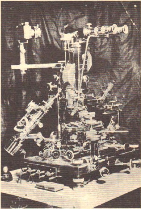
In 1932, Rife received a breast-tumour specimen from the Paradise Valley institution in National City. The initial cultures did not reveal what he was looking for. According to an account later published in a newspaper article by Newall Jones, the turning point came when a culture was left beside an electrically excited argon tube. Rife then observed tiny reddish-purple particles. He designated them BX and regarded them as the virus of carcinoma. The first description of their characteristics in the book is dated 20 November 1932.
More on cultures, experiments and Rife’s interpretations
Lynes states that Rife and his laboratory assistant E. S. Free repeated the procedure one hundred and four times with the same result. Rife’s later account describes four forms: BX; the larger BY, associated with sarcoma; a monococcal form (a form consisting of individual spherical bacteria) in blood monocytes (a type of white blood cell); and a fungal form called Cryptomyces pleomorphia. Rife claimed that they could transform into one another depending on the culture medium. In his report of 1953, he described more than three hundred repetitions of a cycle in which tumours developed after inoculation (the introduction of biological material into an experimental organism) and BX was then recovered from them.
Lynes finds another of Rife’s descriptions particularly telling. A fungal culture derived from the original sample of an unulcerated breast tumor was reportedly kept dormant for a year and then returned to its asparagus based medium. Rife reported finding neither the fungal nor the monococcal nor the BX or BY form, but bacillus coli, which he said would meet ordinary laboratory tests. Lynes reads the story as a striking illustration of pleomorphism: a changed environment supposedly turned the organism into something another observer would classify as an entirely different bacterium. The emphasis rests on Rife’s account of the culture’s origin and transformation. [3]
In the same report, Rife stated that more than four hundred successful animal experiments had preceded use in humans. Lynes presents these statements as the core of the progression from laboratory culture to clinic. He also connects Rife’s explanation with the organism’s internal environment: Rife attributed disease to the effects of microorganisms’ chemical constituents on disrupted cellular metabolism, so his interest extended beyond visible forms to the conditions in which they appeared. [3]
A concluding note to this chapter extends the account to the medium itself. Lynes describes K Medium as a preparation made from dried pig intestine with a saline solution added. In his summary, irradiating the culture supposedly increased the suspected agent’s virulence (the ability to cause disease or more severe illness) and accelerated tumor development in experiments. He then attributes further claims to Rife: that pork and mushrooms provided a natural environment for the organism’s growth and were a natural cause of cancer, and that he had also found it in orchids. The book includes these claims as part of Rife’s broader thinking about media, food and the internal environment supposedly governing microbial behavior. [3]
The frequency instrument complemented the microscope. Rife believed that each microorganism had a characteristic electrical response and that an appropriate frequency could disable it. He called this its Mortal Oscillatory Rate, or MOR. In the newspaper account cited by Lynes, the explanation is compared with the resonance of tuning forks or the response of glass to sound. The book describes a generator, signal amplification, and a gas tube, together with reports of observed organisms disintegrating or ceasing to move. The microscope allowed the experiment to be watched, while the frequency device acted on the specimen. [7]
Microscopes, optics, comparisons and the classification of organisms
In 1933, Rife completed the Universal Microscope, the largest and best-known of his instruments. For the microscope of 1929, Lynes gives a working magnification of 5,000 to 17,000 times; for the Universal, he gives, in the terminology of the time, a “resolution” of 31,000 times and magnification of 60,000 times. Seidel’s description in the book lists 5,682 parts. According to Seidel, two circular, wedge-shaped quartz prisms stood between the light source and the specimen, polarising the light. Further prisms were said to bring the rays back into parallel alignment through the instrument before they could cross. Rife emphasised adjustable monochromatic illumination and the ability to photograph and film the observed activity.
The book presents the full sequence as five microscopes: the 1920 instrument, the second from 1929, the Universal officially completed in 1933, another from 1934, and the last begun in 1937 and finally completed in 1952. Lynes notes that the Universal may already have been used in 1932, while still unfinished, and that later instruments incorporated parts from earlier ones. The sequence is therefore a history of prolonged rebuilding and improvement rather than five entirely separate objects. The final microscope’s beginnings before the war and completion after Rife’s return to work connect two very different periods of his life. [3]
Seidel associated particular microorganisms with characteristic colours: the filterable form of the typhoid organism with turquoise, the filterable form of the tuberculosis organism with emerald green, and BX with reddish purple. Lynes sees this as the instrument’s distinctive feature: it did not merely magnify, but was said to allow living forms to be recognised by their light. In 1933, Rife also wrote Viruses and Rickettsia of Certain Diseases and invited other researchers to examine his observations. Alongside the construction of the microscope, experiments on cultures and animals continued; meanwhile, Johnson was preparing the next step. [3] [7]
The book supplies a later comparison through a demonstration of a smaller Rife microscope before physicians on May 16, 1942. According to the report Lynes takes from the Smithsonian text, a Zeiss ruled grating was viewed first through an ordinary instrument and then through Rife’s. The first reportedly showed fifty lines with chromatic and spherical aberrations; the second showed five greatly enlarged lines filling the field, without such apparent aberrations. An unstained blood film was then compared: the report describes one hundred cells in the ordinary microscope’s field and ten in Rife’s. Lynes uses this description to support his emphasis on magnification and image clarity. [3]
The documentary collection ends with a comparative overview of bacterial and viral characteristics in Appendix X. Beside the names of organisms, the table classifies their motility, changes of form, chemical or polarization properties, length and width, and responses to heat, rays and other conditions. It also lists the colors in which they could supposedly be observed. Alongside ordinary bacterial forms, it includes filterable forms, among them the supposed cancer agent. It thus shows how Rife tried to organize individual observations into a common research system. In the book, this is a historical record of his classification, supplementing the description of the microscope, cultures and frequency instrument. [14]
1933–1934: Johnson opens the way to patients
Johnson’s role in this story was different from Rife’s. He organised visits, wrote letters, approached physicians, and tried to assemble a group that would take the research into a clinical setting. In July 1933, he contacted Karl Meyer, director of the Hooper Foundation in San Francisco. In February 1934, he brought him to Rife’s laboratory.
What Johnson’s letters say about his colleagues’ responses
In letters to Rife and Kendall, Johnson described Meyer’s enthusiasm; Meyer subsequently wrote himself that he was still thinking about what he had seen and wanted to return with his own research material. Lynes later compares these letters with more reserved statements by Meyer and Foord about their involvement. In that discrepancy, he sees an example of former supporters beginning to distance themselves from Rife as the disputes intensified.
In the spring of 1934, Johnson rented space on the Scripps family estate in La Jolla. The book refers to a ranch, a library building, or an annex. In early April, he wrote to Kendall that, beyond the scientific question, he was driven by the thought of people suffering and dying from cancer. He told Rife that he planned to stay in La Jolla from mid-June to mid-September and hoped that, if the work had progressed far enough, they could try the method on a human patient.
Arrangements for the premises and research committee
On 30 April, he invited Rife to inspect the library building before a final agreement was reached on its use. In Lynes’s account, the clinical work is associated with the University of Southern California’s Special Medical Research Committee, led by Johnson. The author also supports the committee’s existence and continued activity with Johnson’s biographical entry in Who’s Who for 1944–1945. The summer clinic thus becomes the centre of a collaboration extending over several years, rather than an isolated event. [3] [4]
Summer 1934: sixteen patients in La Jolla
According to Lynes, sixteen people in very serious condition arrived at the Scripps estate. The book describes them as patients with cancer and other diseases, while Rife’s report describes cases of various malignant conditions. Lynes presents the clinic as the central achievement of Rife’s work: laboratory research was followed by use with patients whom the participating physicians reported as cured.
How the book describes the course of treatment
The book also describes the clinic as a period of learning through the work itself. In its account, the doctors initially repeated the procedures more often and later lengthened the intervals. Rife explained this in terms of an alleged accumulation of breakdown products and the body’s need to remove them. In his later report, he stressed that patients experienced no pain, noise or other perceptible stimulus while the instrument operated; they supposedly saw only a lighted tube. He also wrote that no special diets were used and that no rise in body temperature was observed. Lynes offers these details as a historical description of the first trial’s organization and Rife’s explanation of the reported recovery. [4]
In a report dated 1 December 1953, Rife wrote that, after three months, the medical group had declared fourteen of the sixteen cases clinically cured. He named five physicians and the pathologist Alvin G. Foord; the book also mentions Kendall, Couche, Meyer, and others among those involved. His explanation was that the instrument disabled the microorganism and the body then repaired itself. Lynes connects this report with the physicians’ testimonies and surviving correspondence.
The differing timelines and the case of Tom Knight
The book explicitly describes the remaining two patients as well. In chapter fifteen, it states that they were pronounced cured one month after the clinic closed. In chapter sixteen, it summarises the event with a different timetable: fourteen patients in seventy days and the remaining two in ninety days. The introduction speaks of all sixteen within three months. The book’s main claim is therefore an outcome of sixteen cures, although its accounts of the timing are not entirely the same; here they are retained in the forms in which they appear. [4] [7] [9]
Kendall’s letter of 20 September 1934 singles out Thomas, or Tom, Knight. The lesion on his cheek could be observed and measured, so Kendall recorded its dimensions and details of the examinations. In the letter, he said that he would report Rife’s work to the chairman of the university’s board of trustees. In the same letter, he called the work experimental and described the need for a more powerful instrument. Johnson’s later letters return to Knight: in 1935, he asked Rife to visit him and examine his face, lymph nodes, and lip.
In a statement from 1956, James Couche described a farmer with stomach cancer whom he had seen at the clinic. The man was so emaciated that, on examining him, Couche thought nothing more could be done for him. According to his testimony, the man recovered in approximately six weeks to two months, travelled to his farm in El Centro, stayed awake through the night with a sick cow, and returned. After that visit, Couche wanted an instrument of his own. The book says he used one in his practice for twenty-two years; in the same testimony, he also describes the recovery of a boy with osteomyelitis (a bone infection).
Couche’s account of a boy with a bone infection
Couche’s other detailed recollection concerns a nine-year-old Mexican boy with osteomyelitis of the leg, who wore a splint and used crutches. In the doctor’s memory, his bone had been cleaned every week at Mercy Hospital, leaving him frightened of further pain when he arrived at the office. Couche said he first demonstrated the instrument on his own hand, then used it without removing the boy’s bandage or splint. He claimed that the wound closed in less than two weeks and that the boy later grew into a strong man without a recurrence. Lynes presents the story as Couche’s personal recollection of later use in his practice. [4]
Lynes does not conceal the difficulties with documentation. In December 1935, Johnson explained to Mildred Schram that he had initially conducted the clinic to satisfy himself that the instrument worked in the living body. Without nurses or secretaries, the records were truthful but fragmentary, not prepared for detailed scrutiny by other researchers. Now that he wanted broader support, he acknowledged the need for a different approach to record-keeping.
The author connects the subsequent loss of documentation with Rife’s lending records to Arthur Yale, their later disagreement over changes to the instrument, and the disappearance of university material after Johnson’s death. Despite the gaps, he believes that the letters, the report of 1953, the physicians’ testimonies, and the continuation of clinical work confirm his account clearly enough. His conclusion is that the clinic was an initial experiment with major results, from which more extensive work could have developed with appropriate support. [4]
1935: new instruments, collaborators, and a second clinic
Following Johnson’s comparison, Lynes calls 1935 a year of climbing a mountain. Instrument development, planning a new laboratory, preparations for a second clinic, and negotiations with potential funders were all under way at once. The journalist Newall Jones had learned of the story as early as September 1934, but waited until May 1938 to publish a substantial newspaper account.
An important visitor was the Montreal researcher O. Cameron Gruner. He had obtained a fungal form from patients’ blood on his own asparagus-based medium. According to Lynes, Rife obtained BX from Gruner’s culture on K Medium, and recovered the fungal form from his own BX on Gruner’s medium. Johnson later summarised their experiments as a shared finding that these were different forms of the same organism. He therefore tried to bring Gruner to San Diego for a longer collaboration.
Mrs Young, laboratory work and the development of the instruments
The reproductions of Johnson’s letters also include the case of Mrs. Young. In May 1935, he reported a reduction in lumps in her neck and suggested a further visit. In a September letter, he described Foord’s findings concerning a guinea pig inoculated with material from her lymph nodes: the examination confirmed a tuberculous infection. Johnson then considered Much’s granules as a possible alternative form of the same organism and the need to investigate their response to the instrument. The letter also shows consultation with George Dock and caution about a possible reaction. In the scan used here, the May letter appears twice; it is the same document, rather than two separate testimonies. [14]
Philip Hoyland joined the work on electronics. He collaborated with Rife, Jack Free, and Johnson on a new generator design, completed in September. Johnson’s letters describe the demanding task of matching the new instrument with the old one and the large number of possible settings. The book presents the new designs as part of continuous development aimed at improving the precision, reproducibility, and usability of the instruments.
Rife also built a smaller microscope for wider laboratory use. Johnson estimated the Universal’s price at approximately 30,000–35,000 dollars, while the smaller model was intended to sell for 1,000 dollars or less. In August, E. L. Walker of the Hooper Foundation spent three weeks learning to work with the microscope and cultures. He then continued his research in San Francisco, but, according to the letters cited by Lynes, struggled with a lack of cooperation and tissue samples.
In the first week of November, Johnson opened a second clinic at Santa Fe Hospital in Los Angeles. This time, he kept more careful clinical records. At the same time, he followed some of the patients from 1934, including Tom Knight and Wayne Grayson. At the end of the year, the committee met in Los Angeles. Lynes summarises the year as one of significant progress in instruments, laboratory collaboration, and clinical work, while the crucial question of funding remained unresolved. [5]
1935–1937: funding, testing, and a missed collaboration
The International Cancer Research Foundation in Philadelphia initially seemed to offer the support that would allow Rife to expand his research. In March 1935, it requested information and photographs; in June, Johnson learned that four insurance companies were also interested in funding the work if the foundation approved it. Its secretary, Mildred Schram, visited the laboratory in July, but was in a hurry. Johnson reminded her that Walker had needed three weeks to learn the procedures. Schram then sent mice and demanded identification of the organisms causing their tumours. Johnson replied that their work so far had concerned the organism they associated with human cancer and that the new tasks would represent a substantial departure from the planned work.
Funding correspondence, further tests and clinical reports
In a letter of 15 October 1935, he used the image of climbing a mountain: someone who repeatedly turns back during the ascent and starts along another route may become exhausted before reaching the summit. The foundation’s president, William H. Donner, showed great interest, while Schram insisted on her tests. In December, she announced that the funding decision had been postponed, but abandoned her original demand and instead requested details of the clinic of 1934, endorsed by George Dock’s authority. Johnson again proposed several weeks of work together with a qualified foundation representative. Lynes interprets this lengthy exchange of letters as an example of personal demands and institutional procedures taking precedence over promising research.
Early in 1936, Johnson and Henry Timken arranged the construction of a new laboratory. Its foundations were laid in April, and Rife moved in towards the end of July. Meanwhile, he and Hoyland were redesigning the electronics and developing a new way of generating and studying waveforms. Donner’s visit opened the possibility of a demonstration in Philadelphia. Rife arrived there on 9 May 1936. According to a later letter from Johnson, he was given six or seven tumours without pathology reports (reports on tissue examinations that help identify the nature of a disease). Rife reported that all but three had already been irradiated; from the three remaining specimens, he obtained the characteristic BX using his procedure.
The mycologist (a researcher who studies fungi) Dr Dodge worked with Rife. Johnson emphasised that the two worked together, recorded every step, and both signed the notes each day. Donner’s letter of 22 May, saying that support would not be forthcoming for at least another hundred days or so, therefore disappointed him deeply. He believed a promise of help following a successful demonstration had been broken. He proposed paying for Gruner to work with Rife’s microscope in San Diego, rather than engaging a new researcher in Philadelphia. Donner rejected this on 2 June. The foundation wanted to continue its own tests; when it wrote to Rife again in the autumn, he no longer replied.
Among the individual surviving reports, Lynes also highlights a letter from eye specialist Harry Goodman dated April 28, 1936. It concerned Julia M. Gowdy, whom he had previously examined on March 23. Goodman reported to Johnson a twenty-nine percent improvement in vision in one eye and ten percent in the other over a little more than a month. He also described progress through an everyday task: she had struggled to read the telephone directory but could now find numbers much faster. In Lynes’s account, such letters connect Johnson’s general reports about eye problems with a specific name, examination dates and another physician’s observation. [5]
At home, the work continued. In September 1936, Couche and Jack Free began new clinical work in San Diego. Lynes cites his notes, according to which the first three cancer patients recovered completely. In the same month, Johnson opened a clinic in a home for the elderly in Pasadena. He wrote to Gruner about patients with tuberculosis, carcinoma, ulcers, and other problems, and about results his correspondent would find difficult to believe without visiting in person. He described exceptionally rapid wound improvement to Meyer. In December, news also arrived from Walker in San Francisco: he was said to have independently isolated the fungal form first, and then, from a breast tumour, moving coloured particles that he concluded were Rife’s BX.
Walker’s work was interrupted soon afterward. In April 1937, illness forced him to stop his research; another doctor replaced him but, in Lynes’s account, made no progress. By the middle of the year, the San Francisco work was effectively over. Johnson emphasized that Walker had obtained only five or six tumors from his surgical colleagues in about a year and a half, identifying their lack of cooperation as a major obstacle. Rife also faced health problems at this time: his eyes required another visit to Louisville in May. The attempt at laboratory replication thus ends with illness, limited material and an unsuccessful continuation. [5]
Early in 1937, Johnson and Dock tried, through Charles Martin, to obtain McGill funding for Gruner’s visit. The attempt failed. The committee decided that it must first firmly establish how cancer arose and only then present the treatment publicly. Johnson wanted to avoid premature publicity and false hope; Lynes characterises his caution as a disastrously missed opportunity. Johnson closed the Pasadena clinic on 28 May 1937 and wrote to his colleague Heitger about restored sight in numerous cataract (clouding of the lens of the eye) patients. He spent the summer with Couche in La Jolla, then returned to treating patients in Los Angeles in the autumn. [5]
1937–1938: Beam Ray and the promise of wider production
A commercial route opened in the autumn of 1937. Philip Hoyland moved to San Diego and joined forces with the promoter Hutcheson, the physician James Couche, and Ben Cullen, Rife’s friend from his early years in the city. They founded Beam Ray, with Cullen providing the initial capital. After consideration, Rife agreed on two conditions: the devices must retain the basic principles of his instrument, and every one must be thoroughly tested for its effect on microorganisms before delivery. The book lists fourteen instruments manufactured: two for England, two for Arizona, and ten for physicians in California.
The English order and problems with delivery
In May 1938, B. Winter Gonin, W. V. Blewett, and their colleague Parsons arrived from England. They ordered a microscope, discussed distributing it from London, and bought the first two Beam Ray devices before they had been built. When they arrived in England that summer, their electrical connections were unfinished. Rife had been away from San Diego when they were shipped and had not checked them. Lynes suspects that Hoyland was counting on a paid trip during which he would complete them. Following sharply worded letters, Rife promised help from his associate Henry Siner and planned to follow himself in mid-1939.
Richard Hamer initially rented the third device for Paradise Valley Sanitarium. According to Lynes, he had to remove it when other physicians began losing patients, so he opened a practice in National City. Cullen later described approximately forty treatments a day, two trained operators, and carefully preserved case histories. He particularly remembered an eighty-two-year-old man from Chicago with an extensive malignant lesion on his face and neck. According to Cullen, after several months of treatment only a small dark spot remained, and the patient returned home.
Cullen’s account connects this case with Morris Fishbein, an influential figure in the American Medical Association. Fishbein was said to have learned about the treatment in Chicago and attempted, through representatives from Los Angeles, to acquire a stake in the business. When the offer was declined, Hoyland allegedly received expensive legal assistance. Hoyland believed that his technical contribution entitled him to a larger share than those of the other partners. Lynes presents his subsequent lawsuit as an attempt to take over Beam Ray, backed by AMA interests. In this connection between a business dispute and the association’s influence, he sees the beginning of an organised effort to destroy Rife’s work.
Rife still spoke cautiously in public. In Newall Jones’s article in the San Diego Evening Tribune in May 1938, he declined to claim that he had cured cancer or any other disease. He emphasised the observed destruction of microorganisms, both in cultures and, under certain conditions, in living tissues. Lynes later sets this statement beside the more definite account of 1953, in which Rife described cures at the clinic of 1934. Between the two texts lay failed funding, the breakdown of the business partnership, and a long period during which he had to rebuild his work. [6] [7]
1939: a lawsuit won, a group broken apart
Siner’s visit to a medical centre in New York
In January 1939, on his way to England, Henry Siner visited a medical centre in New York. In a letter to Rife, he described enormous buildings and the surprisingly small space devoted to bacterial research. When he mentioned a microscope said to reveal forms invisible to ordinary optics, he was initially rebuffed. He then showed Kendall’s and Rosenow’s publications; according to his account, the mood changed to great interest. Lynes uses this scene to introduce the contrast between Rife’s small laboratory and powerful medical institutions whose established assumptions he believes Rife threatened.
Rife was preparing to leave for England in May when he received a summons. Hoyland’s lawsuit against Beam Ray came before Judge Edward Kelly on 12 June 1939. The company was represented by the lawyer Bert Comperet. Lynes describes Rife as a man ill-equipped to cope with harsh cross-examination. Cullen remembered his shaking hands, increasingly heavy smoking, and the alcohol with which he tried to calm his nerves. In Cullen’s account, this was where the addiction began that would repeatedly interrupt the laboratory work in later years.
During and after the proceedings, according to Lynes, AMA representatives visited physicians and pressured them to abandon the devices or put their licences to practise at risk. Hamer quickly returned his instrument. Members of Johnson’s committee also withdrew, but Johnson persisted. The author sees the lawsuit as part of the same pressure that was closing practices, breaking up collaborations, and preventing public presentation of the results obtained so far. In this, he draws on associates’ recollections and the research group’s subsequent fate.
Beam Ray won the lawsuit. Kelly denied Hoyland the relief he sought because he did not believe Hoyland had dealt honestly in the business. He expressed confidence in the company’s other directors, while also stating that the court was not judging the device’s value. Lynes writes that the AMA’s alleged role was not examined during the trial, and adds that Kelly reportedly offered afterwards to represent them in a new lawsuit against the association. But Cullen was financially exhausted, had even lost his house, and had to find employment. The company had won, but the group no longer had the strength to continue its planned work.
Gruner’s letter, the question of observation and the laboratory fire
Later in 1939, Gruner wrote to Johnson about a closely related obstacle. He described visiting Rous at the Rockefeller Institute and comparing his photographs with the discussion of “elementary bodies,” which he considered related to BX. After visiting research on the Glover organism in Washington, he concluded that they were observing the same tiny phase, although its development into cocci and bacilli remained an open question. At his own institution, an immunologist and a bacteriologist then checked his work, already prejudiced, he said, against a cancer microorganism. He reported that progress had stalled for nearly nine months. Lynes uses the letter to show the everyday consequences of the larger theoretical dispute. [6]
In the same letter, Gruner strongly supported Rife’s microscope. London opticians had mocked it, he said, invoking the wavelength objection; he thought the main problems were illumination and the observer’s willingness to examine living material. He argued that researchers overlooked particles in their own images that he could recognize. Lynes links this passage with Evelyn Fox Keller’s biography of Barbara McClintock, the 1983 Nobel laureate. From it he draws the themes of patient observation, familiarity with an organism and openness to the unexpected. The comparison helps him explain why, in his view, access to an instrument does not itself ensure that people will see the same thing. [6]
Alongside this story, Lynes recounts the fire that destroyed J. C. Burnett’s research laboratory in New Jersey in the spring of 1939. For fifteen years, the laboratory had studied electrical energy in relation to the human body; the work was funded by Burnett’s wife, Cora B. Timken, a relative of Rife’s supporter. The fire broke out while they were visiting Rife in California. Lynes presents it as an unusual coincidence in the year when the San Diego group also came under its greatest pressure. [6]
The forties: lost support and a microscope without its prism
Even after Beam Ray’s collapse, Johnson continued to seek recognition. In 1942, he sent his instrument to Gruner in Canada. Lynes writes that Gruner left it unused out of fear, and later gave it to a priest and amateur radio enthusiast who used it for radio parts. Couche persisted in San Diego until the mid-fifties; according to the book, this cost him his AMA membership. As late as 1944, Johnson still described himself in his biographical details as the head of the research committee. His death that year deprived Rife of his most important organiser and intermediary.
Suspicions, microscope comparisons and the fate of the instrument
Lynes describes several suspicions surrounding Johnson’s death. A rumour circulated that, shortly before his hospitalisation, he had intended to publish the cancer-treatment results. The author explicitly states that the evidence of possible silencing is circumstantial, then reports that two federal investigators who examined the hospital record in the late fifties or early sixties were said to have considered poisoning probable. The committee’s records were also reportedly destroyed after Johnson’s death. Cullen connected this with the university’s fear of a controversial subject. In Lynes’s account, the loss of the man and the loss of the documentation are two blows to the same work.
Lynes places an earlier wartime episode in 1940–1941, as the electron microscope began to gain ground. In his account, Rife traveled to Germany and compared the new instrument with his own 1929 optics. The author attributes to him an immediate judgment that his own solution better served the goal he pursued: observing living microorganisms. In Lynes’s explanation, preparation for electron microscopy destroys the specimen, removing the possibility of following the same living organism as it changes. He uses the episode to explain the divergence between Rife’s research goal and the subsequent direction of microscopy, which the book connects with different observational habits across an entire generation. [7]
Meanwhile, the microscope was still attracting attention. Raymond E. Seidel began studying it in 1942 and spent three weeks in the laboratory. With his co-author M. Elizabeth Winter, he published an article in the Journal of the Franklin Institute in February 1944; a reprint in the Smithsonian Institution’s annual report followed that same year. He described the complex optics, quartz prisms, and illumination said to distinguish living microorganisms. The paper also mentioned their response to lethal frequencies. According to the book, letters from interested laboratories continued to arrive for several years after publication.
Between 1944 and 1946, a new technician allegedly stole a valuable quartz prism from the Universal Microscope, rendering it unusable. Lynes connects the incident with the publication of Seidel’s article: he sees the article as breaking through the silence and the theft as an attack on the instrument that made further observation possible. Seidel reportedly noticed that he was being followed; later, while he was driving, a bullet pierced his car’s windscreen. The book recounts these incidents as part of a broader intimidation of people who continued to speak publicly about Rife’s discoveries.
Other collaborators were disappearing from the shared work. Through Cullen, Lynes reports that Kendall allegedly received a large payment, withdrew to Mexico, and lost his fortune there. He later lived with his family in La Jolla, where he died in 1959. Rife kept his laboratory until 1946, then began selling it piece by piece. The book describes the combination of alcohol, lack of money, and a research interruption lasting several years, including two years in an institution for treating addiction from which he reportedly eventually escaped. In 1950, he returned to research. This time, John Crane was at his side. [6] [7]
1950–1960: John Crane and a second generation of instruments
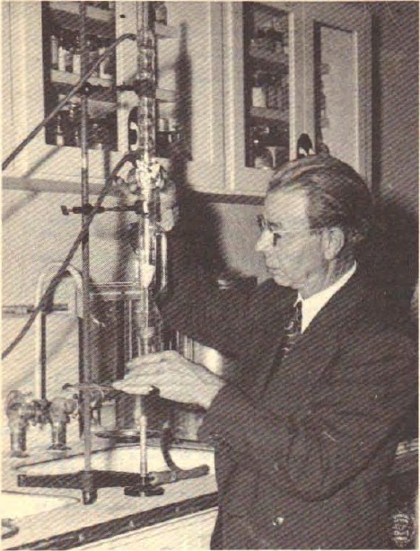
John Crane met Rife in 1950 and decided to devote his knowledge of electronics and mechanical engineering to continuing his work. He obtained one of the original circuit diagrams from Gruner and recruited Verne Thomson, an electronics specialist with the San Diego Police Department, to help build new instruments. Rife did not remain merely a teacher: according to Lynes, he and Crane developed the devices together. Crane also persistently encouraged him to write. The first copyrighted material on the cancer virus appeared in April 1953; the account of the treatment was completed in December and copyrighted the following year.
From 1954 onwards, Crane wrote to the National Cancer Institute and other government institutions. The book sharply criticises a National Research Council committee’s assessment from that year: according to Lynes, it concluded that the discoveries could not work without examining the instruments or consulting Rife, Gruner, and Couche. The author emphasises that government institutions continued to cite this assessment long afterwards. Meanwhile, Crane and Rife carried on with limited resources. Unable to build a large, powerful gas tube, they developed a smaller instrument with body contact between 1954 and 1957.
Rife’s letter and the statements of Tully and Blanche Jones
Rife also recorded his assessment of the later instruments in a 1956 letter that Lynes includes at the end of the chapter on the microscope and frequency instrument. He said that he had worked with the instrument since 1921 and that its form and performance had changed with advances in electronics. He maintained that his many years of research had caused him no injury or other ill effect, and described the equipment as reliable and efficient. He even called the latest model infallible and emphasized its simple operation. Lynes uses Rife’s own testimony to show how highly the inventor regarded development after his renewed start with Crane. [7]
Appendix T contains a statement by dentist Charles F. Tully dated June 1, 1954. Tully described his own prostate inflammation and the case of a friend who, he claimed, had avoided surgery after use of the instrument. He also mentioned lupus with a butterfly-shaped rash, a cancerous lesion that the referring physician considered incurable, and use for postoperative and dental problems. He emphasized that he had known about the device for several years, although his personal use was more recent. He concluded by calling for further research. Lynes includes the statement as signed testimony from a practitioner using the device; these are Tully’s claims about the outcomes he observed. [14]
The appendices also give a patient’s name and the date of sworn testimony concerning Couche’s later work. On January 16, 1968, Blanche H. Jones stated that a breast had been removed because of cancer in April 1956, but that the disease had supposedly persisted after surgery, causing a heavy purulent discharge. According to her statement, James B. Couche treated her with the frequency instrument in May 1956. She claimed that the discharge stopped and that the sarcoma, which she associated with a biopsy in her account, had been cured. She signed the document almost twelve years after the events described. Lynes reproduces it as her personal, notarized statement about the long-term outcome. [14]
In 1957, Crane recruited the physician Robert Stafford of Dayton, Ohio, who wanted to conduct clinical treatments and experiments on mice. After six months, he reported on four cancer patients. In one, he described marked and unexpected improvement. Three terminally ill patients died, but all had experienced relief after treatment began. Lynes also gives the continuation of the report: autopsies were performed on two of them and were said to show another cause of death and surprisingly few cancer cells. Stafford added that he had observed no harmful effects among thirty-three people treated for various conditions.
Stafford’s research collaboration and the assessment of the instrument
In February 1958, Stafford presented his findings to eight physicians on the Montgomery County Medical Society’s general-practice committee. According to the book, they showed interest, and he began assembling a research committee. He even considered manufacturing and distribution in the eastern United States, finding an engineer, a patent attorney, and potential investors. Physicians in Salt Lake City also began using the device, but the local medical board forced them to stop in May 1958. Lynes describes a patient’s bitter disappointment and a physician who told a colleague that he would immediately buy a device for his own family if someone developed cancer.
That same year, a hearing took place before California’s health department. Lynes lists Palo Alto Detection Lab, Kalbfeld Lab, UCLA Medical Lab, and San Diego Testing Lab, and states that all had judged the instrument safe to use. Nevertheless, he reports that the committee headed by public-health director Malcolm Merrill declared the device dangerous and prohibited it. This discrepancy between the described tests and the decision is one of the book’s central arguments that the new period of research was also halted from outside.
According to Lynes, Crane made two further technical improvements in 1958 and 1960. He leased devices under contract, collected reports, and organised training so that users would not alter the design on their own. He needed evidence of usefulness to obtain a patent and investors; to produce more extensive evidence, he needed money and cooperation from physicians. By 1960, he had prepared a manual and distributed ninety instruments for research and testing under notarised contracts. Lynes describes this as a moment of approaching a new breakthrough, followed by another intervention from the authorities. [8]
1960–1965: seizure, criminal proceedings, and prison
In 1960, investigators entered Crane’s office and seized instruments, electronics, and extensive documentation. Lynes estimates the value of the instruments at more than forty thousand dollars. Among the items removed, he lists research reports, technical data, private letters, photographs, invoices, and recordings; he also alleges that the investigators had no warrant. Visits to physicians and individuals testing the devices reportedly followed, along with pressure to abandon the work. The author sees this as the destruction of the research base that Crane and Rife had spent a decade assembling.
Rife, almost seventy-three years old, withdrew to Mexico. The criminal proceedings against Crane took place early in 1961 and lasted twenty-four days. Lynes lists in detail the material that, he says, the defence was not allowed to use: the seized research records, Rife’s statement, medical and scientific reports from the thirties and forties, Stafford’s findings, and Couche’s letters. Crane therefore had to rely mainly on people who had used the devices themselves and were willing to describe their experiences.
Witnesses, the expert and jury selection in Lynes’s account
Fourteen such users testified at the trial. They included seventy-six-year-old James Hannibal, who spoke of his cataracts disappearing, and people who described improvements involving the bladder, intestinal problems, skin lesions, pain, and growths. Lynes singles out the prosecution’s expert, Paul Shea. He reportedly had the device for two months but admitted that he had not tested it or conducted experiments; after inspecting it, he concluded that it had no healing properties. The author also alleges that an AMA physician served as jury foreman, while the other jurors were selected so that they had no medical or electronics knowledge and did not read newspapers sympathetic to alternative treatment.
Crane was sentenced to ten years in prison. Following appeals, the California Supreme Court reversed two of the three counts because specific criminal intent had not been established; he spent three years and one month in prison. These were the criminal proceedings of 1961, separate from the civil lawsuit in which Beam Ray prevailed against Hoyland in 1939. Lynes writes that pressure after the conviction also reached Stafford, who left medicine and became a salesman, and a Salt Lake City physician who died by suicide after sabotage of his device and continued persecution.
After his release, Crane did not give up. In October 1965, on behalf of the Rife Virus Microscope Institute, he again applied to California’s public-health board for approval of the instrument. Rife had returned from Mexico but remained in the background. The reply of 17 November stated that safety and effectiveness had not been demonstrated. Lynes describes a closed circle here: without recognised usefulness, there was no patent or money; without physicians’ cooperation, the evidence demanded by the authorities could not be collected.
Chromiak’s statement and the cases of two chiropractors
Another documentary testimony is the sworn statement of George Chromiak Jr., dated January 7, 1965. He described a Staphylococcus aureus infection contracted in 1960 while working in a Cleveland hospital. According to his account, it became resistant to antibiotics, systemic and chronic, causing prolonged suffering and hospitalizations. He credited the instrument with relief and the disappearance of the systemic infection after years of difficulty. He also reported permanent damage to his right hand and wrist and reduced use of the limb. The statement therefore contains both his belief in the device’s benefit and a description of the lasting consequences of the disease. He concluded by supporting freedom of research. [14]
He illustrates this with two chiropractors. Charles W. Bunner offered a statement about the device’s operation; he was then reportedly visited by the very department that had requested evidence, prohibited from using it, and subjected to a court order for the instrument’s destruction. After submitting his statement, Les Drown was allegedly visited by an American Cancer Society representative who tried to entrap him into an unlawful act; he had to surrender the device or risk imprisonment. For Lynes, these are key examples of his thesis that the authorities were not merely rejecting evidence submitted to them but preventing new evidence from being produced. [8]
Virginia Livingston and the researchers Lynes connects with Rife
Lynes does not look for the continuation of Rife’s story only in electronic instruments. He devotes a substantial part of the book to researchers studying microorganisms whose findings he regards as a belated confirmation of Rife’s direction. Foremost among them is Virginia Livingston-Wheeler, who also published under the names Wuerthele-Caspe and Wuerthele-Caspe-Livingston. According to the book, she began studying tumours in the summer of 1947 and observed the same organism in them. The following year, Eleanor Alexander-Jackson’s work on changes in the tuberculosis bacillus encouraged her to look for similar transitions in the organism she associated with cancer.
Early publications and links between researchers
In March 1948, she presented her findings with the microscopist Roy M. Allen, and in August published them in the bulletin of the New York Microscopical Society. Lynes then connects several lines of research: Gruner’s Canadian work, Couche’s visit in 1949, Irene Corey Diller’s discovery of fungal forms, and James Hillman’s observations at the RCA laboratories. In his account, Livingston’s article of December 1950 demonstrated the transmission of disease to experimental animals and the subsequent isolation of the relevant organism. The author interprets this as fulfilling Koch’s postulates (rules for testing whether a particular microorganism causes a particular disease) and as an important argument for the bacterial origin of cancer that he advocated.
But here, too, Lynes tells a story of closing doors. He attributes the cancellation of Diller’s symposium in 1950 to Cornelius P. Rhoads of Memorial Sloan-Kettering. When Livingston’s group appeared at the international microbiology congress in Rome in 1953, the Washington Post reported on it. According to the book, rejection by the New York Academy of Medicine followed, then the termination of funding for the Rutgers-Presbyterian laboratory, which Lynes likewise attributes to Rhoads. Livingston lost her position and moved to California, first to Los Angeles and, in 1955, to San Diego.
At the congress in Antwerp in 1958, the book says, she found considerably more interest in pleomorphism and immunological approaches. In 1959–1960, she frequently visited Rife’s new laboratory and helped obtain mice for experiments. Their explanations of the changing microorganism were related, but their treatment paths differed: she was developing a serum, he an electrical instrument. Lynes describes this encounter as a connection between two generations that had arrived at similar questions by different routes. Livingston continued publishing and speaking at congresses, while Rife’s associates became embroiled in another court case.
Further experiments, interrupted research and later proposals
Lynes also recounts an experiment that the Milanese researcher Clara Fonti performed on herself in 1959. According to his account, she administered to herself a bacterial culture that the book associates with cancer, subsequently developed a tumor and had it surgically removed. The author presents the episode as a human experiment that, in his view, repeated earlier transfers between blood, cancer tissue, fungal cultures and laboratory animals. This brief report carries considerable weight in his argument: he wants to show that the discussion of the supposed microorganism went beyond observing specimens. [9]
Livingston’s career was interrupted again in 1962 by a heart attack; according to the book, she did little work until 1965. She then prepared a new paper with Alexander-Jackson. In 1966, they spoke at a seminar for science journalists in Arizona, but on returning to Columbia, Alexander-Jackson learned that her work had been stopped. Lynes connects the episode with continuing institutional opposition. In May of the same year, Rife and his colleagues tried to interest Philadelphia’s Institute for Cancer Research in the frequency instrument, but the institution withdrew. For the author, these are two further examples of collaboration being interrupted precisely when broader investigation was being sought. [9]
In the later developments, the book highlights Diller and Florence Seibert, who reported in 1967 that they had isolated bacteria from every tumour they obtained. In 1969, Livingston, Alexander-Jackson, Diller, and Seibert appeared before the New York Academy of Sciences; their paper, published in 1970, described a highly pleomorphic microorganism they associated with various human and animal malignancies. In these descriptions, Lynes hears an echo of Rife, Kendall, Rosenow, and Gruner. He particularly emphasises that the researchers came from different institutions.
Livingston and her husband opened a clinic in San Diego in 1968. According to her figures, summarised by Lynes, by 1983 they had treated more than ten thousand cancer patients and achieved an eighty-percent success rate. In 1972, she published Cancer: A New Breakthrough, followed later by The Conquest of Cancer. Lynes places alongside her publications Lida Mattman’s work on cell-wall-deficient forms from 1974 and Sorin Sonea and Maurice Panisset’s A New Bacteriology. He sees in them the return of questions pushed to the margins in Rife’s time.
Lynes also sets later developments alongside the large publicly funded war on cancer. He states that on December 23, 1971, Richard Nixon signed an act worth $1.6 billion, and that by 1985 the NCI was spending $1.2 billion annually. In her 1972 book, Livingston sharply criticized the expenditure, drug testing and use of patients without their full knowledge and consent. Lynes summarizes her figures of five hundred million dollars over thirteen years, one hundred and seventy thousand drugs tested and more than one hundred thousand patients. He uses these numbers to support his accusation that resources and influence expanded while other research approaches remained unsupported. [9]
Lynes extends her later vision to food and disease prevention. He summarizes Livingston’s warnings about a supposed microorganism in poultry and eggs and her proposal to immunize cattle and poultry with a serum. He then connects this with Rife’s idea of inactivating BX in meat and with his claims about pork and mushrooms. The author also considers treating blood in blood banks. He further attributes to Livingston a proposal for early childhood immunization and the belief that cancer could be eliminated within a decade. The book presents these as historical ideas belonging to her and the author: Lynes uses them to illustrate the scope of a future that he believed the loss of Rife’s work had closed off. [9]
The account also closes a personal circle. In 1976, Livingston married Owen Wheeler, who had known Rife as a young man and spent time in his laboratory. That same year, Christopher Bird published an article about the fate of Rife’s microscope. For Lynes, these two encounters mean that the story had not been broken off despite decades of silence: it survived in people, documents, instruments, and related research questions. [9]
The Victims: the human cost in Lynes’s account
The chapter The Victims interrupts the sequence of laboratories, letters, and court cases. Lynes wants to show the people whom he believes are hidden behind the large death totals. He first introduces Dorothy Lynch of Dorchester, Massachusetts, who sought information about other approaches during her illness. Her experience is chronicled in Exploding into Life, a book in which she and her husband, the photographer Eugene Richards, brought together writing and photographs of illness and hospital life. His second example is nine-year-old Mark Johnson of La Crosse, Wisconsin, author of The Great Planet Swap and Other Stories, who died of leukaemia after four years of illness.
Lynes connects their fate with his central accusation: he is convinced that the suppression of Rife’s work denied people an opportunity that might have changed their lives. The chapter is therefore not simply an account of two individuals, but an expression of his anger and grief. He closes it with a naval officer’s letter to Rife from 1953. The writer recalls visiting the laboratory in his youth, admires Rife’s photomicrographs, and regrets that the world must search anew for answers he believes Rife had already offered. [11]
Cancer and AIDS: the book’s view from the eighties
In the chapter Cancer and AIDS, Lynes extends the account to the epidemic then emerging. His starting point remains Gruner’s idea of an organism that can move between different forms, and the connection he sees between Gruner, Livingston, and Rife. He then introduces the physician and writer Alan Cantwell. Cantwell reported finding the same microorganism in cancer and Kaposi’s sarcoma, which the book associates with AIDS. Separately, he advanced the hypothesis that the same organism also causes AIDS, arguing that AIDS research should take a bacterial explanation seriously. Lynes accepts these assertions as a continuation of the old dispute over pleomorphism.
Gruner’s interpretation of diet and the dispute over research directions
Gruner’s argument in this chapter is more than theoretical. Lynes recounts his claim that a meal containing pork supposedly produced a blood picture indistinguishable from that of a cancer patient for eight or nine hours, after which it returned to normal. He connects this with the pork base of Kendall’s K Medium and with Rife’s observation that such an environment supposedly encouraged the growth of the presumed cancer microorganism. He therefore uses Gruner’s description to extend the argument that the organism changes according to diet or environment. Here the book moves from a laboratory explanation to a much broader interpretation of the relationships between food, blood and disease. [11]
The book sets against this Robert Gallo of the National Cancer Institute, who firmly rejected that explanation. Lynes argues that money went to established directions in research while others were excluded. As another example, he cites a Sloan-Kettering study from 1975: in his account, an organism in blood cultures from cancer patients was explained as external contamination and the work was set aside. For this statement, he directs the reader to Raymond Brown’s book about AIDS, cancer, and medical institutions.
In Aids: The Mystery and the Solution, Cantwell expressed his belief that “AIDS is cancer and cancer is AIDS”. He also speculated about whether some medical procedures had unintentionally contributed to the emergence of more virulent forms of the supposed bacterium. By summarising his claims, Lynes sharpens his warning that, in his view, the story of an overlooked discovery could repeat itself during a new epidemic. This chapter shows how the author understood the urgency of his book in 1987: he wanted not only to recover Rife’s past but to prompt readers to demand a re-examination of his ideas and the opening of other research paths. [11]
1968–1992: final years, surviving documents, and Lynes’s book
In March 1968, a few months before his eightieth birthday, Rife transferred ownership of the microscope to John F. Crane. According to the document described by Lynes, he still intended to patent it, with the rights belonging to Crane. He regarded the frequency instruments as their joint invention, since both had contributed to the new designs. He died in 1971. The instruments, plans, correspondence, photographs, and associates’ memories he left behind became the basis of later efforts to put his work back together.
Crane, the preservation of records and the microscope’s later history
Crane played a central role in preserving this material. Lynes credits his persistence and his refusal to give up when others withdrew. But he also criticises him sharply: Crane lacked Johnson’s organisational skills, drove away capable specialists, businesspeople, and lawyers, and lost years to poorly prepared lawsuits. In 1959, he demanded one hundred and fifty million dollars from potential investors. Later, he filed extensive lawsuits himself against the State of California, Nelson Rockefeller, and many others. Lynes believes that this consumed energy that should have gone into research and collaboration.
Bird’s article What Has Become of the Rife Microscope?, published in March 1976, brought the story back to readers. Between 1984 and 1988, the Universal Microscope passed among groups intending to restore it but, according to the book, they made no progress. A federal court case was ultimately needed to return it to Rife Labs. The company had been established to re-examine the work using modern scientific procedures. The conclusion of the edition used here describes its cautious evaluation of the electronic instruments and the hope that the interrupted research might one day continue.
Barry Lynes met Crane early in 1986. By his own account, he was initially sceptical, but the quantity of surviving documents then convinced him that he must write the story. The first printing of The Cancer Cure That Worked! appeared in March 1987; reprints followed, including the fourth in April 1992, on which this article is based. Mattingly’s foreword calls for Rife’s work to be re-examined and considers how our expectations influence what we are able to see under the microscope in the first place.
Mattingly’s foreword: observation, light and changing forms
In his foreword, Mattingly first considers the observer. People’s senses differ in sensitivity, while knowledge and experience influence recognition of what is seen. Microscopy adds choices of lenses, illumination, stains and specimen preparation. The foreword therefore concerns not only the instrument’s power but also whether a researcher is prepared to recognize an unexpected phenomenon. Through a story about large ships going unrecognized because they lay outside the familiar world, Mattingly illustrates his view that what is obvious to one observer is not necessarily obvious to another. He places this understanding of seeing alongside Rife’s dispute with established microscopy. [1]
The foreword connects Béchamp’s microzymas with Enderlein’s endobionts and Reich’s bions. Mattingly considers them similar, or perhaps identical, tiny living particles observed under different names; he emphasizes dark field microscopy (a method in which illuminated particles stand out against a dark background) and the difficulties of gaining acceptance for unusual methods of observation. He describes Rife’s illumination as even less familiar. He then turns to Gustave Le Bon and later biophysical research into the interaction between light and living systems. His explanation is that adjustable light might tune into a microorganism’s characteristic response and create a resonance that would make it appear to illuminate itself. This is Mattingly’s interpretation of a possible principle behind Rife’s instrument. [1]
Mattingly extends pleomorphism into a broader reflection on life. He connects changes of form with the development of an organism, evolution and symbiosis, challenging the idea that bacteria should be an exception. He uses Béchamp’s explanation to discuss bacteria supposedly originating from particles within living systems themselves, and the relationship between the internal environment, degenerative diseases and pathological forms. In his view, studying dead, stained specimens made it harder to understand processes within a living organism. From these reflections, he argues for a shared framework for observing living specimens and a willingness to reopen old theoretical disputes. [1]
Mattingly is cautious about new microscopes: in his view, ultraviolet light harms the bacteria being observed, while computer enhancement of images may add further complications to interpretation. He mentions a little-known French instrument from the nineteen-sixties, to which he attributes a magnification of 4,500 and a resolution of 150 angstroms (units of length; one angstrom is one ten-billionth of a metre), combining dark field illumination, near-ultraviolet light and a laser. Finally, he describes his own observation of a pleomorphic process in fresh, untreated blood samples using a research microscope and dark field illumination. He emphasizes the importance of prior learning and mentorship. He closes the foreword by calling for a fair re-examination of Rife’s devices and work in the light of new knowledge. [1]
At the end, Lynes urges readers to write to their political representatives and the media. He wants the book to inspire action. The publisher’s closing note places his accusation in relation to particular people from earlier decades, while also expressing respect for the dedication of most physicians and surgeons. The account thus ends with the same hope that links Rife, Johnson, Crane, and the later researchers: that someone would take up the surviving instruments and documents again, examine them, and enable the continuation of work they believed had remained unfinished. [1] [8]
Key milestones in the story
- 1888Rife is born in Elkhorn, Nebraska.
- 1913He moves to San Diego, where he develops his mechanical and optical work.
- 1920–1929He builds his first microscopes and electrical instruments; his work attracts newspaper attention in 1929.
- 1931–1932Collaboration with Kendall and Johnson, the first scientific publications, and a dispute over the interpretation of observations.
- 1933Completion of the Universal Microscope.
- 1934The La Jolla clinic; Lynes reports sixteen cures, while the book gives several different summaries of the timing of the outcomes.
- 1935–1937New instruments, further clinics, collaboration with Gruner, and an unsuccessful search for sustained funding.
- 1938–1939Beam Ray expands instrument production. It wins the civil lawsuit in 1939, but the dispute exhausts those involved and disrupts the business’s momentum.
- 1944Publication of Seidel and Winter’s article on the instrument; Johnson’s death.
- 1950–1957Rife works with John Crane; new designs are developed, including smaller contact devices.
- 1960–1965Seizure of equipment, criminal proceedings, and Crane’s imprisonment; a new application for approval is rejected.
- 1968–1971Transfer of the microscope to Crane in 1968 and Rife’s death in 1971; in parallel, the work of Livingston and other researchers develops.
- 1976–1992Renewed interest, Lynes’s review of the material in 1986, the first edition of the book in 1987, and later reprints.
Sources and notes on the historical material
- Barry Lynes, The Cancer Cure That Worked! Fifty Years of Suppression. Marcus Books, © 1987; fourth printing used here, April 1992, ISBN 0-919951-30-9. Publication details: PDF pp. 2–3; introduction to the contributors and author: printed pp. 1–2 / PDF 7–8; foreword by John W. Mattingly: printed pp. 3–10 / PDF 9–16; early years: chapters 4–5, printed pp. 33–40 / PDF 39–46; publisher’s closing note: PDF 165. PDF numbers refer to the pages of the 166-page scan used here.
- Lynes, chapter 6, printed pp. 41–47 / PDF 47–53: collaboration with Kendall and Johnson, newspaper reports, the dinner in 1931, the Baltimore presentation, and Rosenow’s publications. The book summarises Rife and Kendall’s article in California and Western Medicine (December 1931), the item in Science News (11 December 1931), and Rosenow’s report in Science (26 August 1932).
- Lynes, chapters 7–8, printed pp. 49–58 / PDF 54–63: BX, accounts of experiments, the article from 1933, the Universal Microscope, and Johnson’s letters preparing for the clinic. Microscope photograph: Appendix G, printed p. 149 / PDF 145. Portrait from 1931: Appendix C, printed p. 145 / PDF 141.
- Lynes, chapter 9, printed pp. 59–65 / PDF 64–70: the clinic of 1934, Rife’s report of 1 December 1953, Kendall’s letter about Tom Knight, Couche’s testimony, and Johnson’s letter about the records. Facsimiles in Appendices I–L: printed pp. 151–155 / PDF 147–151. Figures concerning sixteen or fourteen cases and the remaining two: introduction, printed p. 1 / PDF 7; printed p. 61 / PDF 66; printed p. 103 / PDF 104; printed p. 116 / PDF 116. The book’s summaries of the timing differ.
- Lynes, chapters 10–12, printed pp. 67–85 / PDF 71–89: the International Cancer Research Foundation, Gruner, Walker, development of instruments and microscopes, clinics, the laboratory, and funding plans. The letters are summarised from quotations and reproductions in the book.
- Lynes, chapters 13–14, printed pp. 87–99 / PDF 90–101: Beam Ray, the English collaboration, Cullen’s testimony about Fishbein and the AMA, the civil lawsuit of 1939, its consequences, Johnson’s death, and events in the laboratory. Cullen’s more detailed account of Hamer’s patient and Fishbein’s offer: chapter 3, printed pp. 29–30 / PDF 35–36. Allegations and attributed motives are presented as Lynes’s interpretation or the testimonies he cites.
- Lynes, chapter 15, printed pp. 101–107 / PDF 102–108: Kendall’s presentation, microscopy, Rife’s public statement in 1938, the account of the remaining two patients, and Seidel’s description of the optics and frequency instrument. The bibliography, printed p. 139 / PDF 137, lists Raymond E. Seidel and M. Elizabeth Winter, Journal of the Franklin Institute, February 1944, and the reprint in the Smithsonian Institution’s annual report.
- Lynes, chapter 19, printed pp. 127–136 / PDF 125–134: John Crane, new instruments, Stafford’s report and the autopsy statements, laboratory tests, the seizure, the criminal proceedings of 1961, appeals, imprisonment, and the application of 1965. Also Rife’s later years, Crane’s business and legal difficulties, and the microscope’s fate. Photograph of Rife in 1960: Appendix U, printed p. 164 / PDF 161.
- Lynes, chapter 16, printed pp. 109–119 / PDF 109–119: Virginia Livingston-Wheeler, Eleanor Alexander-Jackson, Irene Diller, Florence Seibert, and other researchers whom the author connects with Rife’s work; publications, meetings, and the San Diego clinic. The account of later confirmation of Rife’s ideas follows Lynes’s interpretation in the book.
- Cancer Research UK, Rife machines. Source for the brief contemporary medical note at the end of the article. Web page reviewed on 28 October 2026. Cancer Research UK
- Lynes, chapters 17–18, printed pp. 121–125 / PDF 120–124: The Victims and Cancer and AIDS. The accounts of Dorothy Lynch and Mark Johnson and the author’s extension of his thesis to AIDS belong to the historical and polemical context of the book from the eighties.
- Lynes, chapters 2–3, printed pp. 17–32 / PDF 23–38: Béchamp, Pasteur, pleomorphism, Kendall, and Rivers; the author’s account of American medical institutions, the AMA, advertising, research funding, and private interests. Among others, the book cites E. Douglas Hume, Morris A. Beale, Paul Starr, and Ralph W. Moss.
- World Health Organization (WHO), HIV and AIDS. Source for the brief contemporary medical note. Web page reviewed on 28 October 2026. WHO
- Lynes, documentary appendices N–O, T, V–X: Johnson’s letters about Mrs Young and Foord’s findings (PDF 153–155; PDF 155 repeats the letter on PDF 153); statement by Charles F. Tully dated 1 June 1954 (PDF 160); affidavit by George Chromiak Jr. dated 7 January 1965 (PDF 162); statement by Blanche H. Jones dated 16 January 1968 about treatment in 1956 (PDF 163); comparative summary of bacterial and virus characteristics (PDF 164). John W. Mattingly’s foreword: PDF 9–16. These accounts are presented as testimony and interpretations in the book.
A brief contemporary medical note
This article presents Barry Lynes’s historical account. Cancer Research UK states that there is no reliable evidence that Rife devices treat cancer. AIDS results from advanced HIV infection. Rife devices must not replace or delay established medical care. [10] [13]